A simple wearable patch for continuous real-time cardiac imaging? A team specializing in ultrasound is working on this.
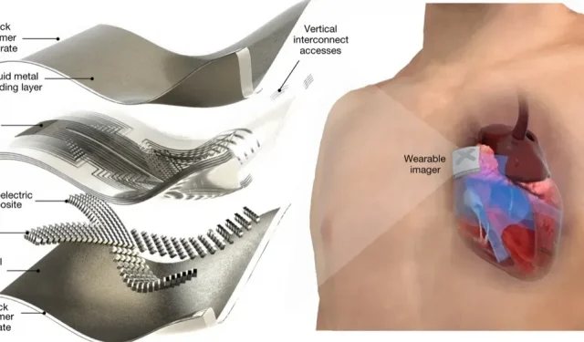
Ultrasounds can provide detailed images of your heart, but the sheer size of the devices makes them unsuitable for continuous monitoring, let alone out of the hospital. However, this technology may become much more portable in the relatively near future. Researchers have indeed developed an ultrasound patch that allows real-time imaging of the heart, even when you’re moving. The system uses deep learning to automatically calculate ventricular volume and generate performance statistics. This way you will know, for example, your cardiac output at all times.
A simple wearable patch for continuous real-time cardiac imaging?
The device uses piezoelectric transducers to image deep tissues. Meanwhile, the stretchable liquid metal electrodes ensure that the accessory stays close to your skin while remaining very compact. Previous attempts at portable ultrasound used very thin metal films, which limited the complexity of the overall design.
However, this technology is very far from being implemented on the market. The scientists still want to miniaturize their system, a system that currently has to be connected to an external processing unit with a flexible cable. The team also hopes to improve spatial resolution with better algorithms and use an AI training dataset to better reflect the world’s population.
A group of ultrasound specialists is working on this
Such a system already has very obvious advantages. The creators are convinced that this can provide continuous data on patients with heart problems or in critical condition, including in outpatient settings. Remote ultrasound scanners have been introduced before, but one had to go through devices that were cumbersome to say the least. This technology may also be useful for athletes who hope to strengthen their heart and optimize their performance.
And this concept is not limited to this one body. The researchers explain that their portable ultrasound system can be adapted for use on the spine, liver and veins. The technology could then provide freedom of movement for many patients and athletes who would otherwise have to travel to specialized facilities for testing.


